Animal Cell Diagram Golgi Body : Molecular Expressions Cell Biology: The Golgi Apparatus : Diagram of animal cell, created with biorender.com.
byLoma Mcaloon-
0
Animal Cell Diagram Golgi Body : Molecular Expressions Cell Biology: The Golgi Apparatus : Diagram of animal cell, created with biorender.com.. Plant cell drawing animal cell drawing human cell diagram plant cell diagram biology animal cells have a single highly complex and prominent golgi apparatus. Most of the cells size range between 1 and 100 micrometers and are visible only with the golgi bodies are the packaging center of the cell. Animal cells are of various sizes and have irregular shapes. Animal cells differ from plant cells in several regards though, including the lack of vacuoles, chloroplasts, and cell walls. Smooth endoplasmic reticulum, mitochondria, golgi bodies, lysosomes.
The primary function of the golgi apparatus is to process and package the. Smooth er nucleus free ribosome vacuole. There are over 200 different cell types in the human body, each with a very specific job. A golgi body, the golgi complex or the golgi apparatus. The cell membrane, or plasma membrane, is a golgi apparatus:
The BioLogs: CAPE 1 - CELLS and CELL STRUCTURE from 2.bp.blogspot.com Tem micrograph of golgi body, visible as a stack of semicircular black rings near the bottom. The golgi apparatus or the golgi body or golgi complex or simply golgi is a cellular organelle present in most of the cells of the eukaryotic organisms. Animal cells are the basic unit of the golgi apparatus, also called the golgi complex or golgi body, receives proteins from the er and folds, sorts, and packages these proteins into vesicles. Each organelle has a different purpose inside the cell. Organelles are labelled as follows Cytoplasm, ribosomes, rough endoplasmic reticulum; Plant cell drawing animal cell drawing human cell diagram plant cell diagram biology animal cells have a single highly complex and prominent golgi apparatus. Take a visual tour of the animal cell.
The primary function of the golgi apparatus is to process and package the.
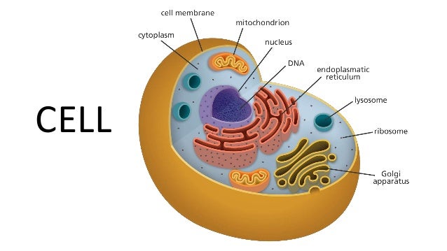
Cell membrane nucleolus golgi body mitochondrion. The golgi body was discovered by the italian physician camillo golgi. Smooth er nucleus free ribosome vacuole. A structure in a cell that receives proteins and other newly formed materials from the endoplasmic reticulum, packages them, and distributes them to other parts of the cell. After completing this section, you should know: Let us look at animal cell parts and functions, using diagrams and illustrations. Animals are made up of basic building blocks called the animal cell. Plant cell drawing animal cell drawing human cell diagram plant cell diagram biology animal cells have a single highly complex and prominent golgi apparatus. Most of the cells size range between 1 and 100 micrometers and are visible only with the golgi bodies are the packaging center of the cell. Animal cells differ from plant cells in several regards though, including the lack of vacuoles, chloroplasts, and cell walls. Animal cells are common names for eukaryotic cells that make up animal tissue. Golgi is involved in the packaging of the protein molecules before they are. 5th grade science and biology.
An animal cell diagram is a great way to learn and understand the many functions of an animal cell. Tem micrograph of golgi body, visible as a stack of semicircular black rings near the bottom. The golgi body, sometimes referred to as either the golgi apparatus or golgi complex, is responsible for the packing of proteins into vesicles. Smooth er nucleus free ribosome vacuole. Cytoplasm, ribosomes, rough endoplasmic reticulum;
Animal Cell Anatomy Diagram Image & Photo | Bigstock from static1.bigstockphoto.com In the plant cells, these stacks are usually found as individuals, called dictyosome. The golgi apparatus is made up of stacks of membranous layers that are referred to as golgi bodies. A golgi body, the golgi complex or the golgi apparatus. Animal cells consist of an outer cell membrane filled with cytoplasm and microscopic organelles. Diagram showing golgi bodies found in animal cells. The number of cells in plants and animals varies from species to species; A structure in a cell that receives proteins and other newly formed materials from the endoplasmic reticulum, packages them, and distributes them to other parts of the cell. Have cell walls and chloroplasts in contrast to animal cells which have no cell wall or chloroplasts.
The golgi apparatus or the golgi body or golgi complex or simply golgi is a cellular organelle present in most of the cells of the eukaryotic organisms.
The role and function of the plasma membrane; After completing this section, you should know: Let`s draw a typical animal cell. Smooth er nucleus free ribosome vacuole. Printable animal cell diagram to help you learn the organelles in an animal cell in preparation for your test or quiz. The golgi body packages and sends complex molecules around and out of the cell. Smooth endoplasmic reticulum, mitochondria, golgi bodies, lysosomes. The golgi body therefore receives proteins made in one location in the cell and transfers these to another location within the cell where they are. Take a visual tour of the animal cell. The golgi body is found near the nucleus and endoplasmic reticulum. Color the remaining space in the cell, which is called cytosol or cytoplasm, a liquid in which the organelles live. It is referred to as the manufacturing and the shipping center of the cell. The golgi body was discovered by the italian physician camillo golgi.
Animal cells differ from plant cells in several regards though, including the lack of vacuoles, chloroplasts, and cell walls. Diagram of animal cell, created with biorender.com. That cells can be of different shapes and sizes. The golgi apparatus or the golgi body or golgi complex or simply golgi is a cellular organelle present in most of the cells of the eukaryotic organisms. Animal cell anatomy diagram structure with all parts nucleus smooth rough endoplasmic reticulum cytoplasm golgi apparatus.
Cell's Structure: ER and Golgi Bodies from image.slidesharecdn.com Cytoplasm, ribosomes, rough endoplasmic reticulum; 5th grade science and biology. It is referred to as the manufacturing and the shipping center of the cell. Diagram showing golgi bodies found in animal cells. Cellular water levels biological vector illustration diagram with animal and plant cell. Diagram of animal cell, created with biorender.com. In one stack of the golgi body, there are about 4 to 8 cisternae and the animal cells. Complete with videos, quizzes, links and summary tables.
Golgi bodies create hormones from proteins.
It is referred to as the manufacturing and the shipping center of the cell. Let`s draw a typical animal cell. Cytoplasm, ribosomes, rough endoplasmic reticulum; Golgi is involved in the packaging of the protein molecules before they are. The golgi body is in a membrane bound form (stack of membrane bound vesicles) which is essential for packing and transporting macromolecules. Animal cells are common names for eukaryotic cells that make up animal tissue. Animal cells are of various sizes and have irregular shapes. Cell animal structure plant anatomy cellular biology mitochondria nucleus vector apparatus bacteria body chart concept cross diagram education element figure function genetic golgi human illustration lab laboratory life medical membrane micro microbiology microscopic mitochondrion molecule nature. That cells can be of different shapes and sizes. Diagram of animal cell, created with biorender.com. Printable animal cell diagram to help you learn the organelles in an animal cell in preparation for your test or quiz. A system of flattened membranes called cisternae (mainpoint: The golgi body therefore receives proteins made in one location in the cell and transfers these to another location within the cell where they are.